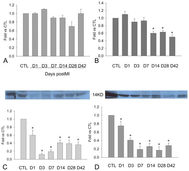
Figure 3.
Cardiac PDGF-B expression: Compared to controls, PDGF-B mRNA remained unchanged at the border zone (penal A), while levels decreased in the infarcted myocardium after week 2 (panel B). PDGF-B protein levels at the border zone (panel C) and infarcted myocardium (panel D) were significantly reduced for over the course of 6 weeks.

