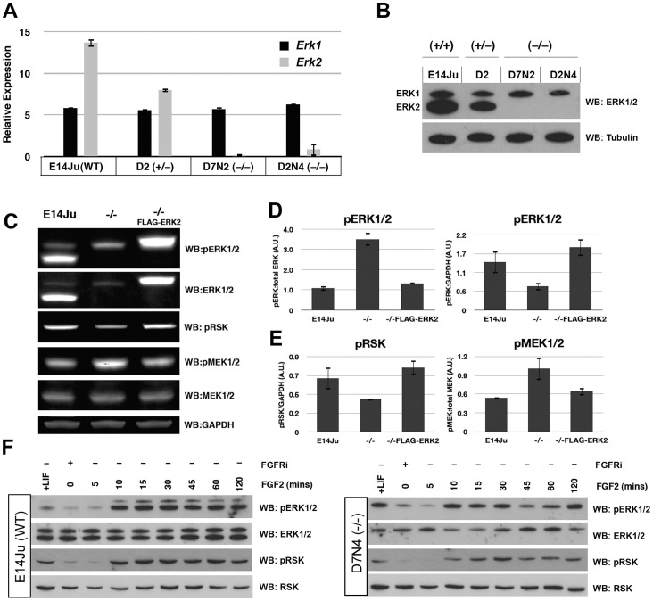
Figure 2. ERK2 ablation attenuates FGF-ERK signaling.
(A) qRT-PCR analysis showing reduced or absent Erk2 transcript in the mutated lines, with no compensatory increase in Erk1 expression. (B) Western blot analysis of total ERK1/2 protein levels in WT, heterozygous, and homozygous Erk2 cell lines. ERK2 expression is reduced in the heterozygous line D2, and completely absent in the Erk2 −/− lines D7N2 and D2N4. Tubulin is used as a loading control. The qRT-PCR and western results for the heterozygous line D7 were identical to clone D2 (not shown). (C) Western blot analysis of wild-type (E14Ju), Erk2 −/−, FLAG-ERK2-rescued Erk2 −/− ES cells. The levels of phospho-ERK1/2, phospho-RSK and phospho-MEK1/2 were examined in self-renewing culture conditions. (D) Quantification of western blotting data to examine the ratio of phosphorylated ERK1/2 to total ERK1/2 (left), and pERK1/2 to GAPDH (right) in E14Ju, Erk2 −/−, and ERK2-rescued Erk2 −/− lines. (E) Quantification of western blotting data to examine the ratio of phosphor-RSK to GAPDH (left), and phospo-MEK1/2 to total MEK1/2 (right) in E1Ju, Erk2 −/−, and ERK2-rescued Erk2 −/− lines. (F) Western blot analysis of ERK1/2 and RSK activation in wild-type (E14Ju) and Erk2 −/− ES cells following acute FGF2 stimulation. Phosphorylation of p90RSK is reduced in the absence of ERK2. Membranes were stripped and sequentially blotted with the indicated antibodies.

