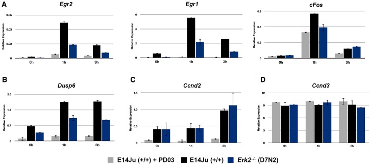
Figure 3. Attenuation of immediate early gene (IEG) induction in ERK2-deficient ES cells.
(A) Analysis of IEG induction by qRT-PCR for Egr2, Egr1, and cFos following acute stimulation with FGF2. WT (E14Ju) and Erk2-null (D7N2) ES cells were cultured for 24 h in the presence of the FGFR inhibitor PD173074 (PD17). The inhibitor was then washed out (0 h), and cells were stimulated with (10 ng/ml) recombinant FGF2 for 1 and 3 hours. To determine basal MEK-ERK independent expression of the genes, WT cells were stimulated with media containing both FGF2 and the MEK1 inhibitor PD0325901 (PD03) (E14Ju+PD03) (B–D) FGF2 induction results for the ERK1/2 phosphatase Dusp6 (B), the late response gene Ccnd2 (C), and Ccnd3 (D). Error bars represent the standard deviation from the mean of biological replicates.

