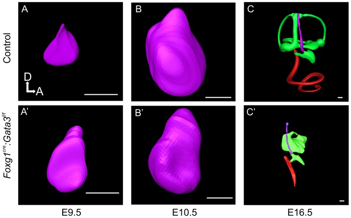
Figure 2. Morphological development of Foxg1Cre∶ Gata3f/f ears.
3D reconstruction of Foxg1Cre:Gata3f/f inner ears compared to control; utilizing confocal microscopy and Amira software. A-C) At E9.5 there is very little difference in size between the two genotypes. By E10.5 there is a slight reduction in the mutant ear dorsally. At E16.5 there is morphologic development of a cochlear duct (red) in the mutant ear, although it is truncated compared to control. There is also morphologic development of the vestibular portion of the mutant ear (green) although it is highly abnormal. While there is a noticeable saccular out-pouching, none of the other vestibular structures are easily identifiable. The endolymphatic duct is present in the mutant ear (purple) and extends dorsally. Dorsal is up anterior is to the right in all images. All scale bars represent 100 µm.

