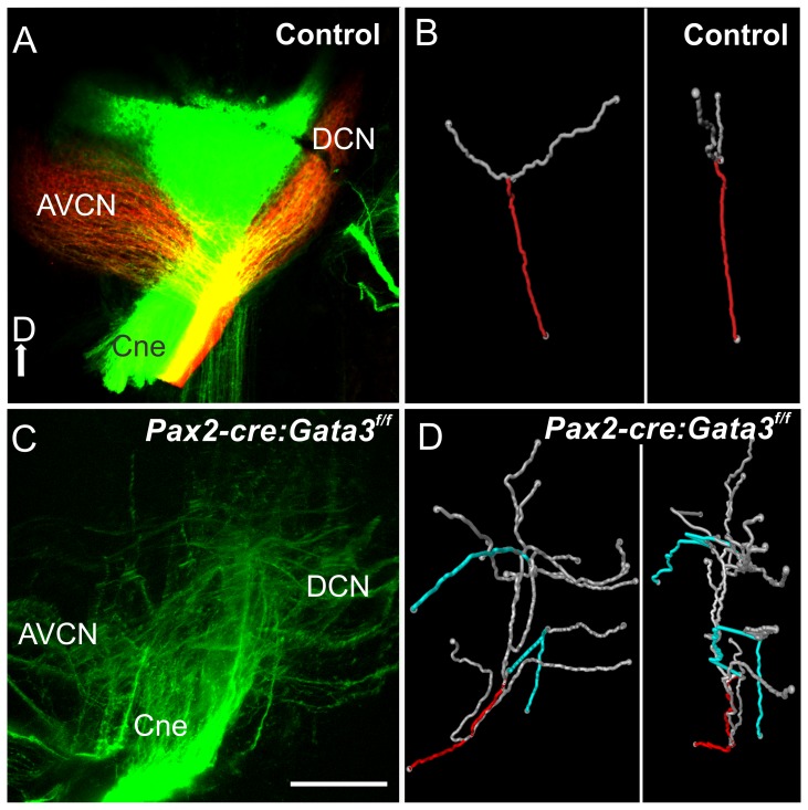
Figure 7. Inner ear central projection.
Lipophilic dye was placed into the cochlea of E18.5 mice. In the control mouse red and green dyes were placed into the base and apex. A and C) Confocal image of cochlear nerve (Cne) projection into the hindbrain (cochlear nucleus). In the control there is a single bifurcation of each afferent axon to the dorsal cochlear nucleus (DCN) and antero-ventral cochlear nucleus (AVCN). In the mutant the cochlear fibers are bifurcating at several branch points with the terminal fibers looping and misdirected. B and D) Amira software was used with the confocal images from A and C to trace individual axons. A single axon was reconstructed, and the root of this axon is labeled in red, and its branches in gray. B) A representative control axon. Each axon analyzed had a single bifurcation. D) In the mutant blue branches are neurites looping back toward the entry point. The single neuron has many bifurcations compared to the control. The right panel of B and D) shows the fibers rotated 90 degrees. The control shows the fibers staying within a single plane. The mutant fibers do not show this restriction in 2D space and branch throughout the dorsal-ventral extent of the hindbrain.

