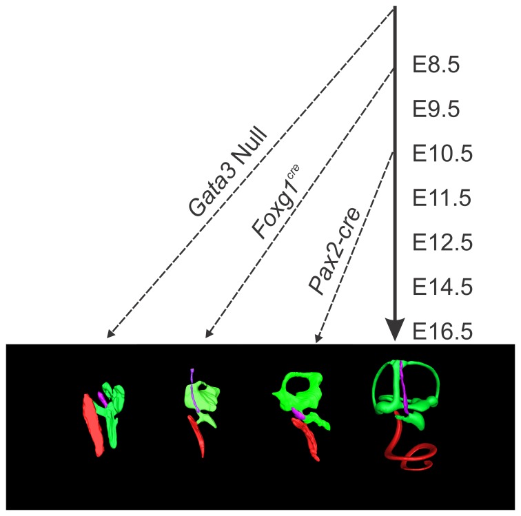
Figure 8. Timing of Gata3 loss reveals different phenotypes.
The loss of Gata3 during different stages of development reveals changing roles of Gata3 in cochlear neurosensory development. In this diagram the solid arrow indicates normal embryonic progression while the dashed arrows indicate the aproximate embryonic age of Gata3 loss from the ear and subsequent altered development. In the Gata3 null mutant [38] the inner ear was shown to contain only a single patch of sensory epithelia (saccule) and corresponding afferent neurons. The cochlear duct was completely devoid of all neurosensory cell types. In the Foxg1Cre conditional Gata3 mutant there is formation of saccule, utricule, and anterior canal neurosensory cell types. The cochlear sensory epithelia is not properly specified, some prosensory genes remain, and many sensory differentiation genes are absent or downregulated. Only in the Pax2-Cre mutant where recombination occurs later than the other two indicated muants (between E8.5 and E10.5) are some cochlear neurosensory cells present, but this later loss of Gata3 results in improper differentiation of these cell types.

