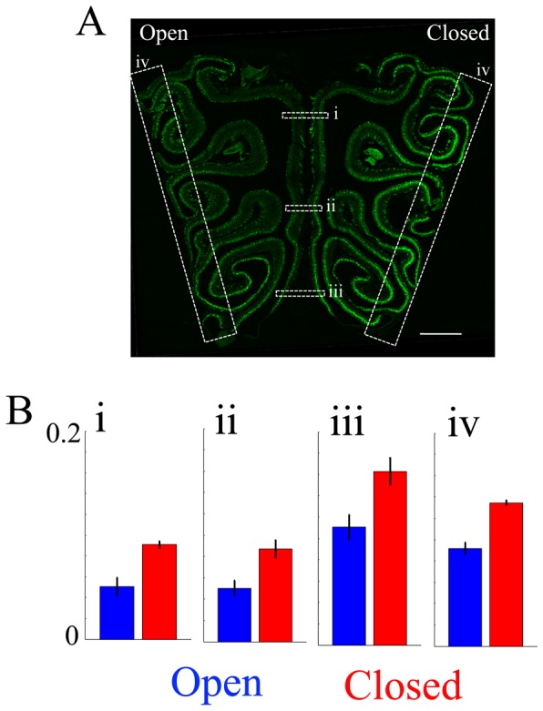
Figure 1. Naris occlusion upregulates GFP immunofluorescence in the occluded side of the OE of TRPM5-GFP mice.

A. This panel shows a representative image of a 14 µm coronal section of the OE taken from a naris-occluded animal. The right side in the image is ipsilateral to the occluded naris (Closed) (scale bar = 500 µm). GFP immunofluorescence is green. B. Averaged GFP immunofluorescence (intensity ranges from 0 to 1, with a gain set so that OSN GFP immunofluorescence ranged from 0 to 0.2). The left side in the image is ipsilateral to the open side of naris (Open). Averaged GFP immunofluorescence intensity in the OE in the septum (i–iii) and the lateral regions (iv) were compared between open and closed sides. Averaged fluorescence intensity was significantly higher in the closed side of epithelium in all locations. (i; p = 0.02, ii; p = 0.006, iii; p = 0.002, iv; p = 0.007, p value FDR corrected 0.05, paired t-test, n = 4).
