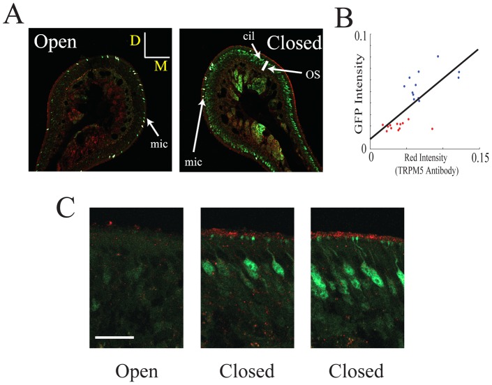
Figure 2. Naris occlusion upregulates the intensity of ciliary layer immunolabeling with a TRPM5 antibody.
Immunolabeling for TRPM5 (red) and GFP (green) is shown in the naris open and occluded sides under two different magnifications. A. Immunolabeling in the open and closed nostrils in endoturbinate II (bar is 50 µm, D is dorsal and M is medial). Notice that there is more intense labeling for both GFP and TRPM5 in the olfactory epithelium soma layer (OS) and cilia (cil) respectively in the closed nostril and that within each turbinate the labeling was inhomogeneous (some areas in the section show higher intensity than others). In addition, as expected from our earlier study [27] there is intense labeling of microvillar cells that are not being studied here (mic). B. GFP immunolabeling intensity in the olfactory soma layer vs. TRPM5 immunolabeling intensity in the ciliary layer (red) measured in 20 degree sections around the endoturbinates in A (see the methods). Intensity in the image ranged from 0 to 1. Blue: closed, red: open. The straight line is a best fit for all the points. The correlation coefficient is 0.72 (different from zero, p<0.0001). The intensity for both GFP and TRPM5 immunolabeling in the open nostril are statistically significantly different compared to intensity in the closed nostril (t-test, p<0.000001). C. Higher magnification figures for the TRPM5 (red) and GFP (green) immunohistochemistry (bar is 20 µm, no microvillar cells are found in these images). The two images for the epithelium in the closed naris were from areas that displayed different intensity for labeling for the two antibodies.

