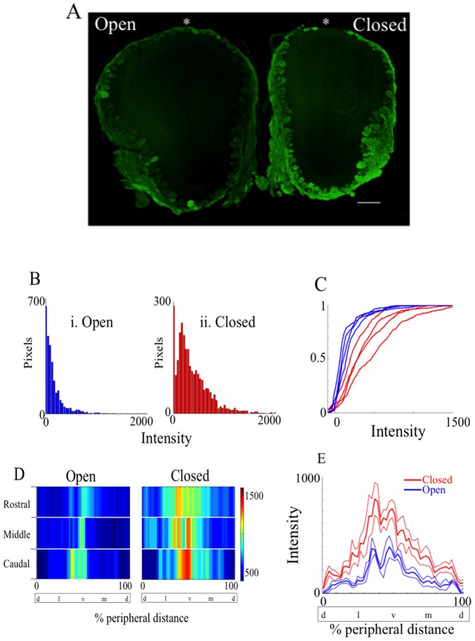
Figure 3. Naris occlusion upregulates GFP presence in the OB.
A shows the representative coronal 18 µm section of the naris-occluded OB of a TRPM5 GFP animal (scale bar = 500 µm). The right OB in the image is ipsilateral to the occluded (Closed) naris, and the left OB is ipsilateral to the open naris. The section was immunostained with an antibody against GFP (green). As expected, the OB is smaller in the occluded side [52]. (B) Histogram of the number of pixels as a function of the fluorescence intensity (0–4095) after subtracting intensity taken from the external plexiform layer (EPL) just underneath the glomerular layer. GFP immunofluorescence is higher in the occluded side (ii) compared to open side (i). (C) Cumulative histogram for fluorescence intensity after subtraction of EPL fluorescence levels for all four animals examined. Occluded OBs (red) express GFP at significantly higher level than open OB (blue) (t-test for mean intensity, p = 0.0286, n = 4). D shows a 2D color map of the glomeruli displaying GFP immunofluorescence as a function of percentage distance from the dorsal most point (*) around the glomerular layer in the olfactory bulb in A. Three representative OB sections were taken from the rostral, medial and caudal one-third and analyzed for GFP immunofluorescence intensity around the glomerular layer. (E) Mean fluorescence intensity around the glomerular layer of the occluded (red) and the open OB (blue). Thin lines represent the standard error of the mean (SEM). The intensity was averaged for caudal, middle and rostral images. Occluded side of OB (red) significantly differs from the open side of the OB (blue) (p<0.0001, N-Way ANOVA, n = 4). % peripheral distance was measured starting from the dorsal most point. d = dorsal, l = lateral, v = ventral, m = medial.

