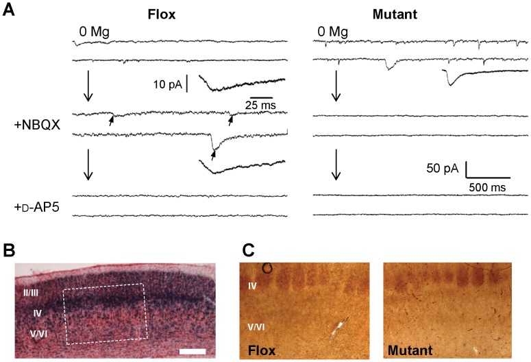
Figure 2. No NMDA currents detected in most of cortical layer II/III pyramidal neurons and postnatal onset of GluN1 deletion.
A. An example trace (right) showing no NMDA currents were detected in mPFC layer II/III pyramidal neurons. Nine cells out of 10 cells tested from 7 mutants (12–17 weeks old) showed no NMDA currents, while they (arrows) were always detected in fGluN1 (Flox) controls. To isolate the NMDA component of spontaneous EPSC events, 20 µM NBQX was bath-applied, and, furthermore, 50 µM D-AP5 was added to ensure that NMDA channel currents were blocked. Scale bar is 500 ms and 50 pA for long traces and 25 ms and 10 pA for average excitatory currents. B. X-gal staining of the primary sensory (S1) cortex on postnatal day 6 (P6) of G35-3-Cre/R26lacZ mice. C. Immunohistochemistry staining for 5-HTT on P6 revealed no difference in barrel field development in the S1 cortex of fGluN1 (Flox) control and mutant mice. Scale bar: 200 µm.

