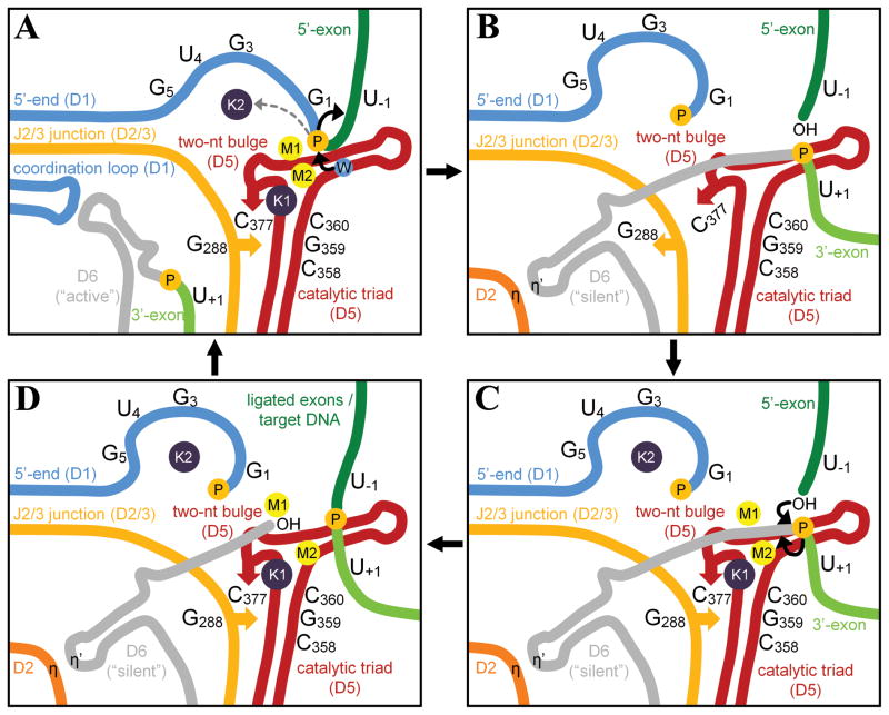
Figure 7. A model for group II intron splicing.
A) Precatalytic state. Group II intron adopts the triple helix conformation and catalyzes 5′-exon hydrolysis. “W” is the nucleophilic water molecule. B) Transient intermediate. Upon hydrolysis, the concerted rotation of G288 and C377 disrupts the metal center to favor the release of G1 and the recruitment of the 3′-splice junction, while D6 in the “silent” conformation forms the η-η′ interaction with D2 (Pyle, 2010). G288 interacts with residues near the bulge in D6 (de Lencastre et al., 2005). C) The second step of splicing. After all reactants are rearranged correctly, the metal cluster forms again to release ligated exons and the free intron. The position of M2 in the second step of splicing remains uncertain, as described in the text. D) SER and retrotransposition. The free intron harbors a fully assembled cluster and can rehydrolyze ligated exons or reinsert into genomic DNA. The retrotransposed intron is then transcribed again to start a new splicing cycle. See also Figure S6.

