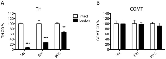
Figure 2. Successful dopaminergic lesions do not cause changes in the COMT protein levels.

A normalized optical density (OD) analysis of (A) tyrosine hydroxylase (TH) or (B) COMT positive neurons in the substantia nigra (SN) and striatum (Stri) after a unilateral 6-OHDA (8 µg) lesion of the SN, and TH and COMT positive neurons in the prefrontal cortex (PFC) after 6-OHDA lesion of the VTA. At least 3 separate stainings were done, and mean ± SEM are shown. Significant decrease in TH OD was seen in all areas analyzed but none in COMT OD. ***, P<0.001; **, P<0.01 vs. control side (Student t-test).
