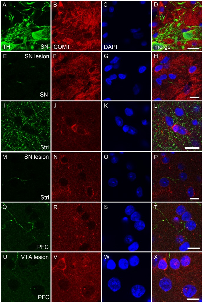
Figure 4. A–X. COMT protein neither colocalized with tyrosine hydroxylase (TH) positive neurons nor decreased after destruction of dopamine neurons.
The effect of dopaminergic lesion on the COMT immunoreactivity. The 6-OHDA lesion of the substantia nigra (SN) and ventral tegmental area (VTA) were made as described and COMT immunoreactivity analysed in the projection areas. In the intact SN, COMT (red color) was not colocalized with dopaminergic neurons (tyrosine hydroxylase, TH; green color; A–D), and lesion in the SN did not affect COMT immunoreactivity (E–H) although TH-immunoreactivity disappeared (E). The situation was similar in the striatum (Stri; I–L versus M–P). Moreover, in the intact prefrontal cortex (PFC), the projection area of the VTA, COMT did not colocalize with dopaminergic nerves (Q–T). Moreover, the lesion in the VTA did not effect on COMT on the PFC (U–W). Nuclei are visualized by DAPI (blue color). Scale bars are 10 µm in all pictures. Three separate stainings were made and representative examples are shown. Quantification of ODs was done in Fig.2.

