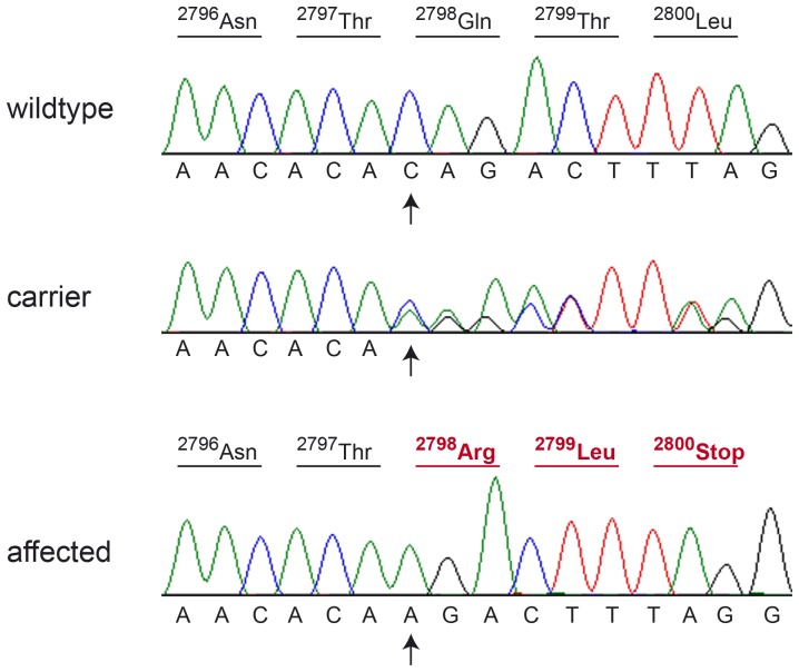
Figure 2. Sanger sequencing of the CUBN:c.8392delC variant.
Electropherograms of a homozygous wildtype, heterozygous, and homozygous mutant dog, respectively, are shown. The position of the deletion is indicated by arrows. The predicted amino acid translation is shown above the sequence. Altered codons in the affected dog are shown in red. The deletion results in an early premature stop codon (p.Q2798Rfs*3).

