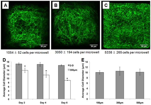
Figure 3. Confinement of hESCs in 3-D microwells leads to a reduction in cell size.
Cells in 300 μm/side microwells were fixed and immunolabeled with an antibody against membrane protein E-cadherin on day 2 (A), day 4 (B) and day 6 (C) of culture. The images show qualitatively that cell size decreased with increasing culture time as the microwells filled. At the same timepoints, cell diameter was measured in cells harvested from Matrigel-coated TCPS 2-D controls and 300 μm/side microwells (D). While cell diameter remained unchanged in the 2-D controls, hESCs cultured in microwells were smaller than those in 2-D controls by day 2, and cell diameter continued to decrease over time. Cell diameter was also compared in three microwell sizes (100, 300, and 500 μm/side) and there were no statistically significant differences in cell diameter between microwell sizes (E) (* indicates P<0.05 compared to same-day 2-D control).

