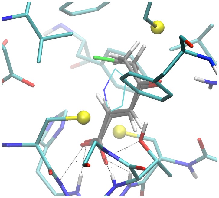
Figure 6. Close view of the docking pose of BrOxoPA and OxoPa in configuration 4.
Enzyme residues are displayed in “licorice” with CPK colors and Carbons in cyan. Small spheres highlight sulfur atoms. Same representation with carbon in grey for inhibitor docking pose. Atoms forming bonds with the carboxylate of PYC in 1W62 structure are connected to corresponding pose inhibitor atoms. PYC molecule modeled by superposition of carboxylate and C2 atoms on that of BrOxoPA is displayed in lines, carbons in cyan.

