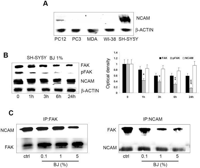
Figure 9. BJ effects on association between FAK and NCAM.
(A) Western blotting analysis of NCAM basal expression in PC3, MDA-MB231, WI-38, PC-12 and SH-SY5Y cells. Note that NCAM is expressed only in both SH-SY5Y and PC12 cells. (B) Western blotting analysis of FAK, pFAK, and NCAM expression in SH-SY5Y cells treated with 1% BJ for different times (from 1 h to 24 hs). Relative densitometric analyses of immunoreactive bands normalized to the values for β-actin are presented in the lateral histogram (mean ± SEM of three experiments). *P<0.05, **P<0.01 and ***P<0.001 vs untreated cells. A slight reduction of NCAM within 6 hs of BJ incubation accompanied to a significant decrease of pFAK is shown. (C) Protein extracts from SH-SY5Y cells treated for 24 hs with 0.1–5% BJ were subjected to immunoprecipitation with FAK antibody (IP:FAK, left panel), or with NCAM antibody (IP:NCAM; right panel) and then analysed for the expression of both NCAM and FAK.

