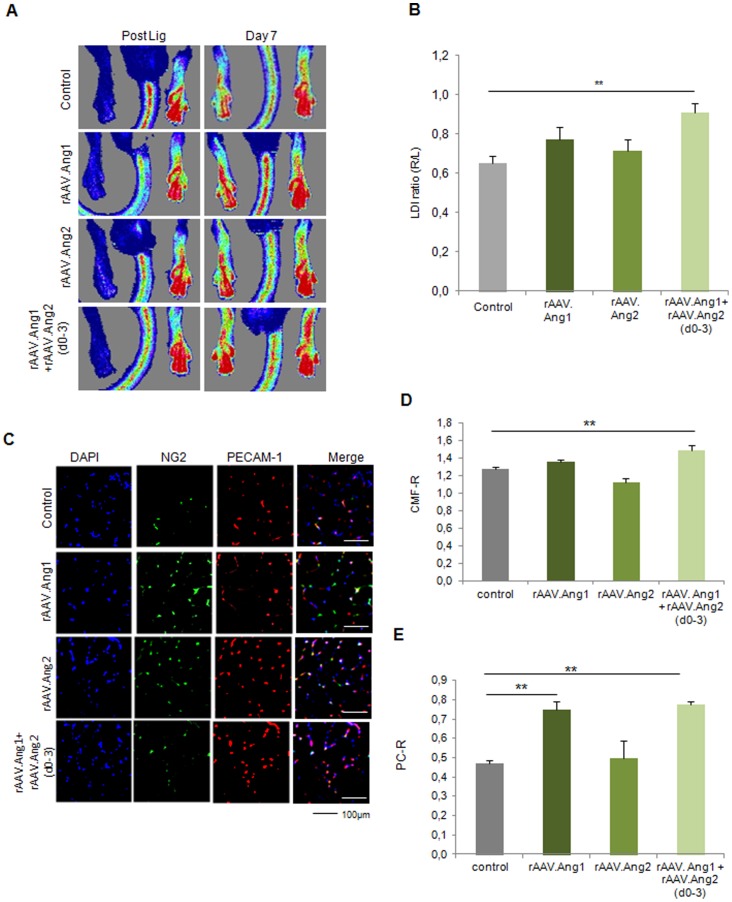
Figure 6. Ang1 combined with early Ang2 (d0-3) induces enhanced neovascularization at d7.
(A, B) LDI analysis revealed no alteration of perfusion at d7 by either Ang1 or Ang2 (continuous overexpression), but a significant increase in the Ang1+Ang2(d0-3) group. (C–D) Analysis of capillaries revealed an elevated CMF-R only in the Ang1 and Ang2(d0-3) group, whereas the capillarization was unaltered if Ang 1 and Ang2 were overxpressed alone. (C,E) Ang1 alone as well as Ang1+Ang2(d0-3) was capable of enhancing pericytes coverage compared to control, Ang2 alone had no effect on the pericyte/capillary ratio (PC-R). (MEAN ± SEM, n = 7,* p<0.05, ** p<0.01).

