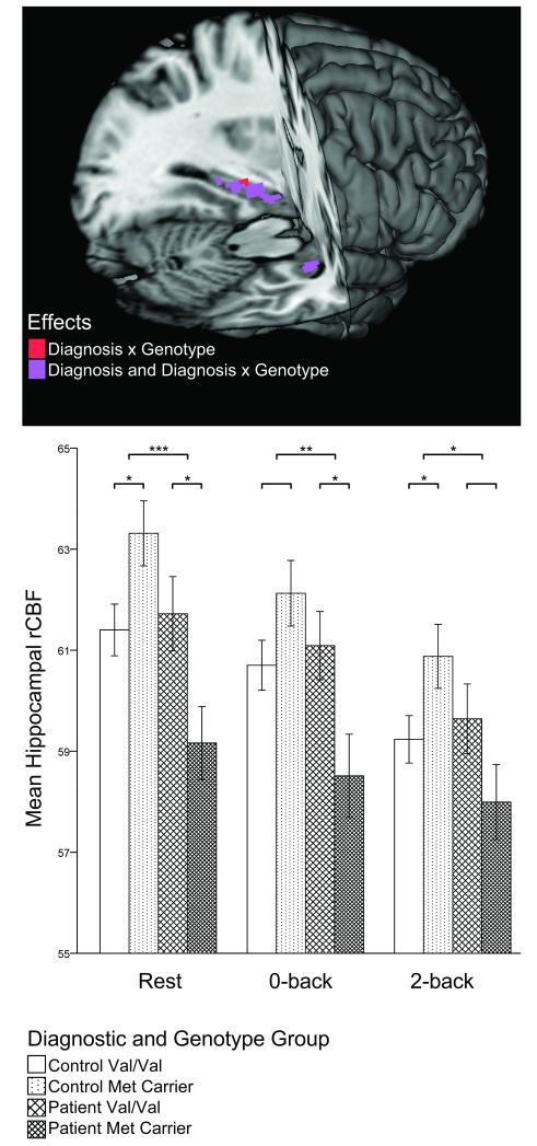
Figure 1.
Scaled Mean Hippocampal Regional Cerebral Blood Flow (rCBF) by Diagnosis and BDNF Val66Met Genotype During Rest, Sensorimotor (0-back), and Working Memory (2-back) Conditions.
In the lower panel, error bars represent standard errors of the mean. Significant within diagnostic group genotype comparisons (lower brackets) and significant diagnosis-by-genotype interactions (upper brackets) are indicated with asterisks (* p≤0.05; * p≤0.005; *** p≤0.001). The upper panel reveals localization of results for voxel-wise resting rCBF comparisons. The search volume was restricted to the hippocampal ROI, and a voxel-wise FDR corrected threshold of p<0.05 was used.

