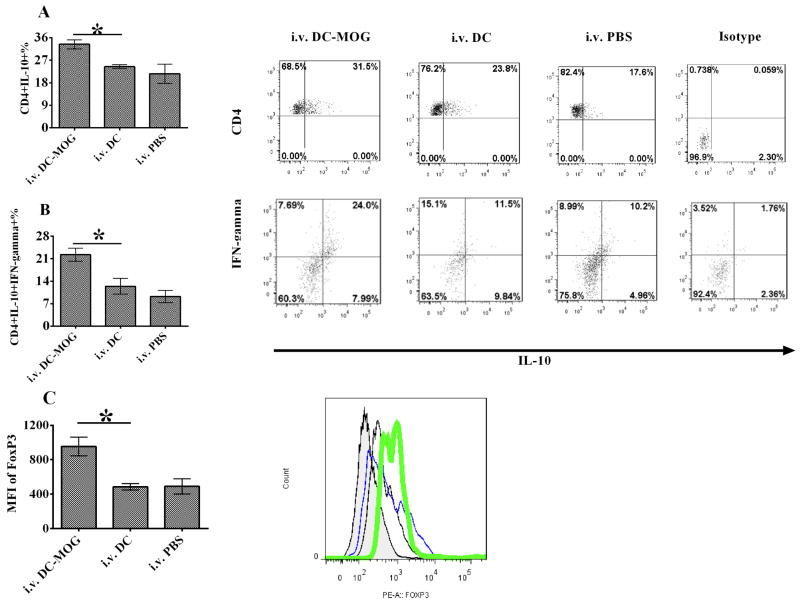
Figure 2.
Intravenous transfer of bone marrow-derived DCs up-regulates the numbers of IL-10 producing CD4+ T cells, suppressive CD4+ IL-10+ IFN-γ+ T cells and FoxP3+ expression in CD4+ T cells. EAE mice were treated with i.v. PBS and DCs pulsed with MOG peptide at 0.1 μM (DC-MOG) or not (DC). Splenocytes were isolated from mice and stained. CD4+ T cells were gated. The production of IL-10 in CD4+ T cells (A), CD4+ IL-10+ IFN-γ+ (B) and expression of FoxP3 (C) in CD4+ T cells were determined by flow cytometry. (C) Expression of FoxP3 in CD4+ T cells is improved after i.v. transfer of DC-pulsed with MOG peptide. Expression of FoxP3 was detected in CD4+ T cells which were isolated from mice with EAE treated with MOG-pulsed DCs (green), DCs without loading MOG peptide (DC) (blue) or PBS (black) as described in Fig. 1. Isotype control is also shown (shade). Error bars shown in this figure represent the mean and SD of triplicate determinations of the percentage of CD4+ T cells (A and B) or mean fluorescence of intensity (MFI) (C) in three independent experiments (n=3, t test, * P<0.05).

