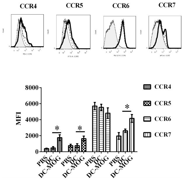
Figure 4.
Intravenous transfer of bone marrow-derived DCs pulsed with MOG peptide up-regulates protein expression of CCR4, CCR5 and CCR7 on CD4+ T cells. Splenocytes were isolated from mice treated with PBS (thin line), DC spulsed with MOG peptide (thick line) or DCs without loading MOG peptide (dash line). The isotype (shade) controls are also shown. CD4+ T cells were gated and their expression of CCR4, CCR5, CCR6 and CCR7 was determined by flow cytometry. Error bars shown in this figure represent mean and SD of mean of fluorescence intensity (MFI) of CCR4, CCR5, CCR6 and CCR7 expression on CD4+ T cells in three independent experiments ( n=3,t test, *P<0.05).

