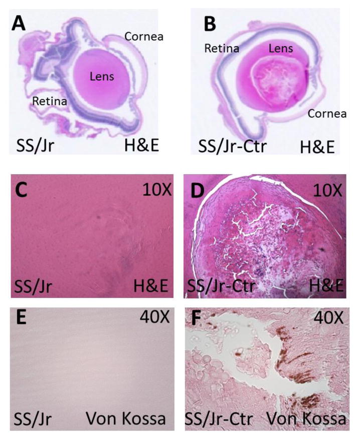
Figure 3. Representative whole-eye and high resolution images of lens from SS/Jr and SS/Jr-Ctr at 21 days of age.
(A–B) Longitudinal histology section through whole-eye (1X, H&E) which demonstrates the normal appearance of key anatomical structures in each strain, aside from obvious lens fiber changes observed in eye from SS/Jr-Ctr. (C–D) Higher magnification images (10X, H&E) showing normal lens appearance in eye from SS/Jr and disruption of lens fibers in eye from SS/Jr-Ctr. (E–F) Lens from SS/Jr-Ctr also demonstrates distinct calcification (40X, Von Kossa stain), whereas wild-type SS/Jr have normal appearance.

