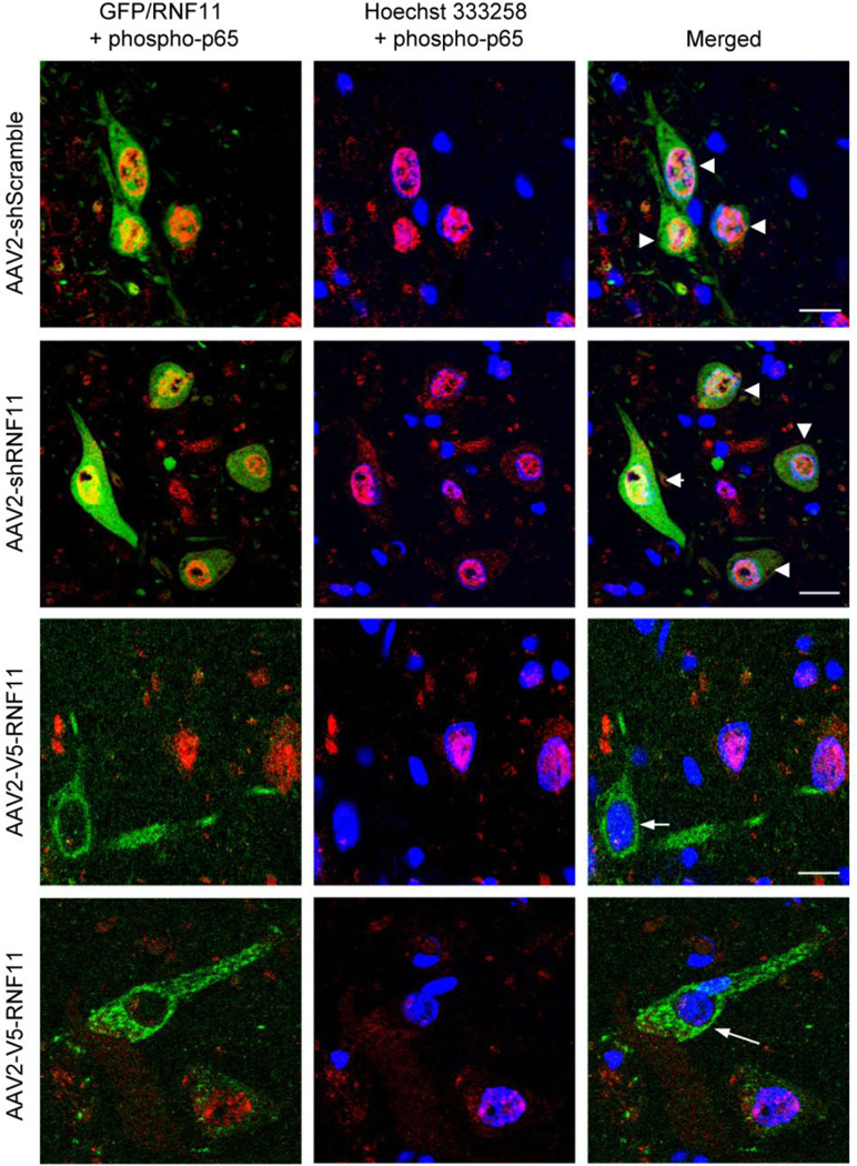
Figure 4. Validation of RNF11 as a negative modulator of NF-κB signaling in vivo.
Representative images of virus expression (GFP or RNF11, in green) and phospho-p65 (in red) at the level of the substantia nigra in AAV2-injected hemisphere. Note the colocalization (arrowheads) of Hoechst 333258 (in blue) and phospho-p65 in the AAV2-shScramble and AAV2-shRNF11 animals (first and second rows) and minimal colocalization in AAV2-V5-RNF11 animals (third and fourth rows) in virus-positive nigral neurons as indicated by white arrows. Scale bar: 20 µM.

