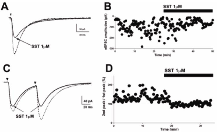
Fig 2.

SST inhibits presynaptic glutamate release. (A), Representative averaged peak eEPSC before (dotted trace) and after (solid trace) SST application reveals diminished synaptic potency of eEPSCs following SST application. (B), Time course change in eEPSC amplitude following SST application in cell shown in (A). (C), Representative averaged eEPSCs obtained by paired stimuli before and after application of SST show that SST application abolished paired-pulse facilitation. (D), Time course change in the paired-pulse response following SST application. Dotted lines at 100% indicate the normalized amplitude of eEPSCs evoked by the 1st stimulus.
