Fig 6.

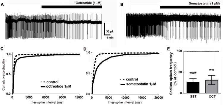
Octreotide and SST reduced the firing rate of CA1 pyramidal neurons. (A, B), Representative trace of a loose-patch recording from CA1 neurons in ACSF containing elevated potassium before and after the application of octreotide (A) or SST (B). (C, D), Cumulative probability plots of neuronal firing before (- - -) or after (—) octreotide (C) or SST (D). (E), Bar graph demonstrating the significant decrease in neuronal firing rate following the application of octreotide or SST.
