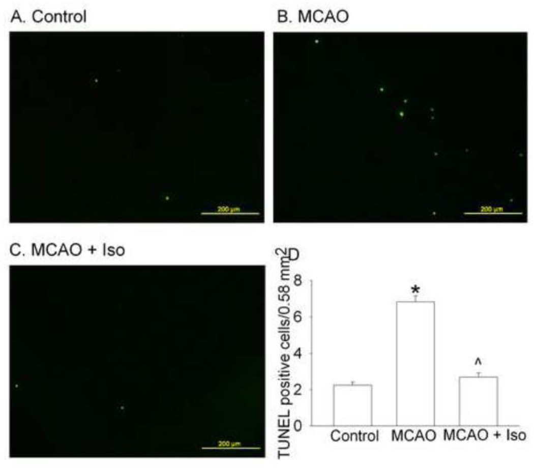
Fig. 3. Isoflurane postconditioning reduced dying cells evaluated at 4 weeks after the 90-min right middle cerebral arterial occlusion (MCAO) in rats.
Cells in the penumbral cerebral cortex were examined after terminal deoxynucleotidyl transferase-mediated biotinylated UTP nick-end labeling (TUNEL). Panels A, B and C are representatives of the sections from control, MCAO only and MCAO plus isoflurane postconditioning. Panel D: quantitative data are presented as the means ± S.E.M. (n = 5 for the control group, = 17 for MCAO, and = 18 for iso + MCAO groups). * P < 0.05 compared with the control group. ^ P < 0.05 compared with the MCAO only group. Iso: isoflurane postconditioning.

