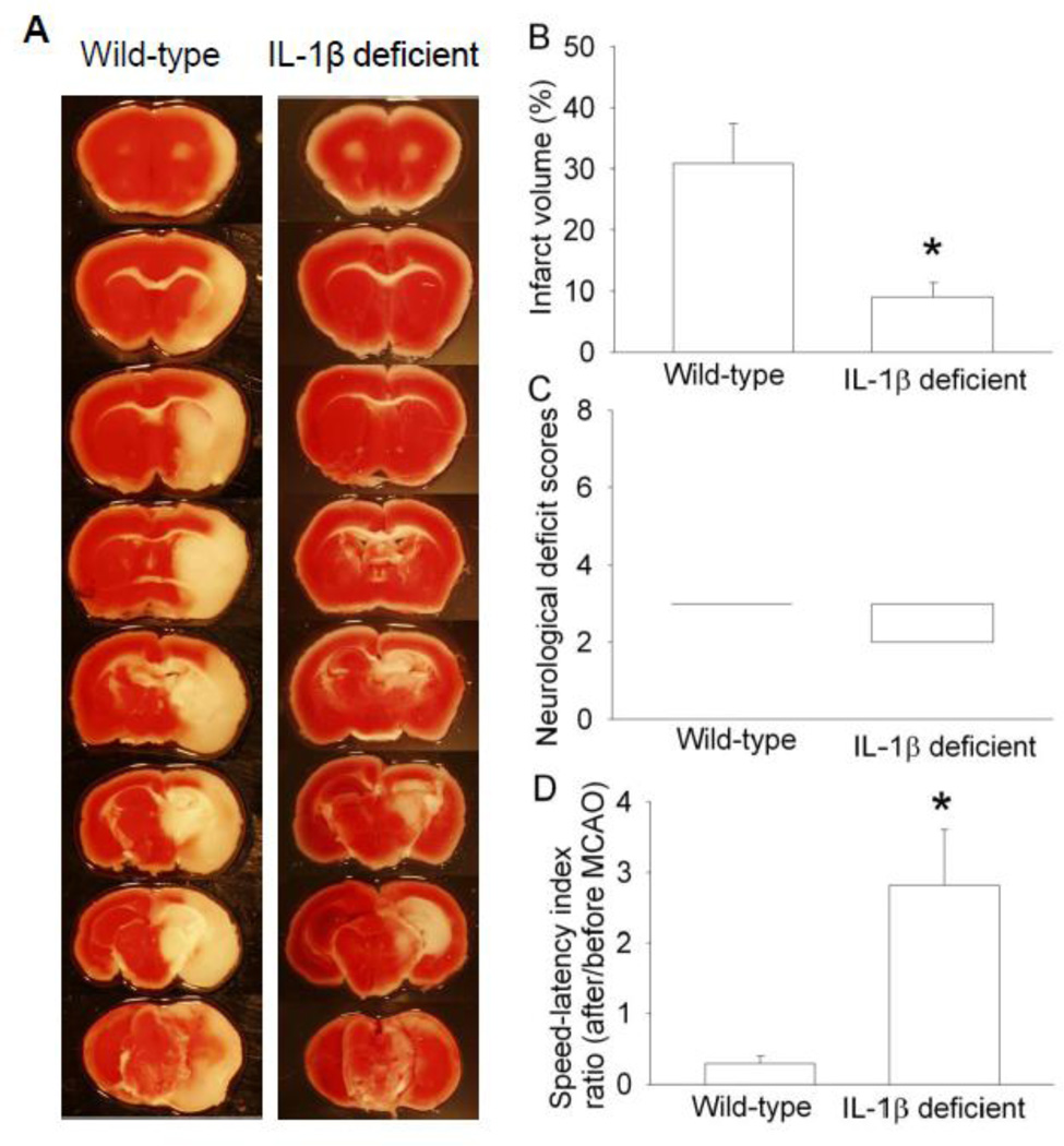
Fig. 7. Interleukin (IL)-1β deficient mice had better histological and neurological function outcome than wild-type mice after middle cerebral arterial occlusion (MCAO).
The results were evaluated at 24 h after the MCAO. A: Brain slices stained with 2,3,5-triphenyltetrazolium chloride from representative mice. B: Percentage of infarct volume in ipsilateral hemisphere volume. Results are the means ± S.E.M. (n = 6 – 8). C: Neurological deficit scores evaluated immediately before the animals were euthanized for the assessment of infarct sizes (data are presented in panel B) or assigned 7 to the animals that died before the end time point for observation. Results are presented in a box plot format (n = 6 – 8). ●: lowest or highest score (the score will not show up if it falls in the 95% interval); between lines: 95% interval of the data; inside boxes: 25–75% interval including the median of the data. D: The performance on rotarod. Mice were tested before and 24 h after the MCAO and the speed-latency index ratio of these two tests are presented. Results are the means ± S.E.M. (n = 6 – 8). * P < 0.05 compared with the wild-type mice.

