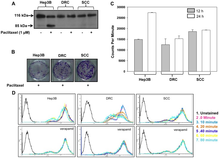
Figure 2. Selection and characterization of paclitaxel resistant DRC and SCC clone.
(A) Hep3B cells, DRC and SCC treated with or without 1 µM paclitaxel for 48 h and PARP cleavage was analyzed by immunoblot. (B) 3×10⁴ cells were plated in 12 well plates for 24 h followed by treatment with 5.2 µM paclitaxel for 48 h. Medium was replaced with drug free medium and incubated for additional 18–20 days. Cells were then fixed and stained with crystal violet. (C) 1×10⁵ cells were plated in 12 well plates. After 24 h, ∼500 nCi/well of 3H paclitaxel was added to each well for 12 h and 24 h, respectively. Cells were lysed by adding SDS to respective plates. Counts were taken on Packard. (D) Hep3B cells, DRC and SCC (106/ml cells) were loaded with Rhodamine-123 (2 µM) for 30 min at 37°C and efflux at respective time interval was measured by MOFLO in the presence or absence of verapamil.

