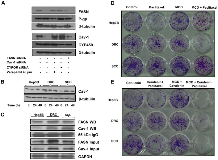
Figure 8. Cav-1 knockdown inhibit the expression of FASN and vice versa.
(A) DRC (5×10⁵) were plated in 35 mm petri plate. After 24 h, cells were transfected with siRNAs targeting Cav-1, FASN or CYPOR as per manufacturer instruction. Simultaneously, 40 µM verapamil was added for 24 h. Thirty-six hours posttransfection or 24 h verapamil treatment, cells were harvested and lysates were prepared. Fifty microgram whole cell lysate proteins were resolved on 8% or 10% SDS-PAGE and western blot was performed. (B) Hep3B cells, DRC and SCC were treated with 300 nM paclitaxel for 24 and 48 h respectively. Whole cell lysates were prepared and 30 µg was resolved on 10% SDS-PAGE and western blotting was performed. (C) Co-immunoprecipitation of Cav-1 and FASN in Hep3B cells, DRC and SCC was carried out using FASN specific antibody. Cav-1 and FASN were detected in the immune complex by immunoblotting. IgG heavy chain and GAPDH served as loading control. (D and E) Hep3B cells, DRC and SCC were plated and allowed to adhere for 24 h and cells were pretreated with MCD (4 h) or cerulenin (24 h). After inhibitor treatment, paclitaxel was added for additional 48 h. Cells were washed with PBS, fresh medium was added and cells allowed to form colonies for ∼ 21 days. Colonies were stained with crystal violet and photographed.

