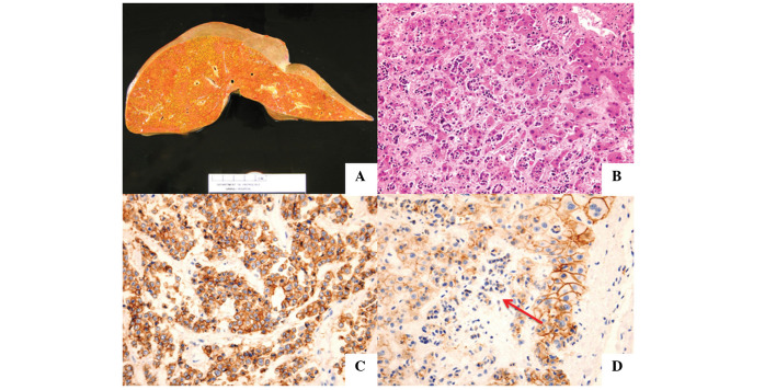
Figure 1.
(A) Image of the unfixed liver revealed hepatomegaly without gross metastasis. (B) Microscopic examination of the liver revealed diffuse infiltration by pleomorphic cells with some glandular formation. (C) Immunohistochemical staining for E-cadherin expression in the metastatic nodule in the lung was strongly positive, (D) that of metastatic tumor cells in the liver was negative (arrow).

