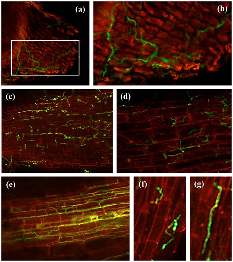
Figure 1. Early stages of chickpea root infection by Fusarium oxysporum f. sp. ciceris races 0 and 5 in compatible and compatible interactions.
(a, b) germinated conidia on the root apex with primary mycelia at 1 dai; (c–d) primary mycelia and initial hyphal colonization on lower root zone at 2 dai; (e) intermediate root zone showing hyphal colonization with mycelium extending from the epidermis into cortical tissues at 6 dai; (f) conidia on the root surface with germ tube(s) at 1 dai; (g) germinated conidia on the root surface with primary mycelia at 1 dai. The Fusarium oxysporum f. sp. ciceris isolates were transformed with the ZsGreen fluorescent protein and imaged using confocal laser scanning microscopy. dai: number of days after inoculation.

