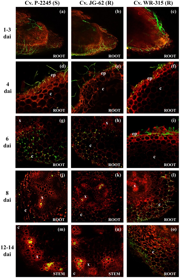
Figure 2. Temporal and spatial patterns of chickpea infection by Fusarium oxysporum f. sp. ciceris race 0.
Colonization of three cultivars, including P-2245 (susceptible, S), JG-62 (resistant, R), and WR-315 (R) was imaged using confocal laser scanning microscopy. ep: epidermis, c: cortex, x: xylem, dai: number of days after inoculation. (a–c): root apex; (d–i): lower root zone; (j): upper root zone; (k): intermediate root zone; (l): lower root zone; (m): stem fifth internode; (n): stem fourth internode; (o): half root zone. The Fusarium oxysporum f. sp. ciceris isolate was transformed with the ZsGreen fluorescent protein.

