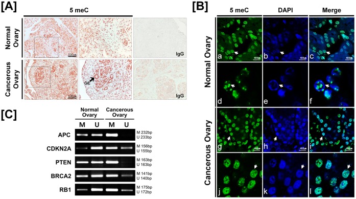
Figure 2. Methylation patterns of DNMTs and tumor suppressor genes in normal and cancerous ovaries from laying hens.
[A and B] Localization of 5-methylcytosine protein in normal and cancerous ovaries of hens. Sections were not counterstained. Arrows in panel B indicate nuclei in the glandular epithelium of ovaries. [C] Methylation status of promoter regions of tumor suppressor genes using methylation-specific PCR analyses. Legend: GE, glandular epithelium; M, methyl primer; U, unmethyl primer. See Materials and Methods for a complete description of the methods.

