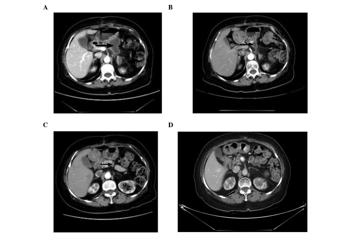
Figure 1.
(A) November 10, 2010: Postoperative arterial phase contrast-enhanced computed tomography (CT) scan showing a low-density nodule. (B) February 14, 2011: Following 4 cycles of CIK cell infusion, the arterial phase contrast-enhanced CT scan shows that the low-density nodule has markedly decreased in size. (C) July 11, 2011: Following 12 cycles of CIK cell infusion, the arterial phase contrast-enhanced CT scan reveals that the low-density nodule has slighlty decreased in size. (D) February 29, 2012: The CT scan shows that the low-density nodule has almost completely disappeared.

