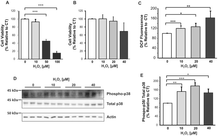
Figure 1. Establishment of mOS conditions for treatment of mouse primary cortical cultures. Cortical cells were treated for 6 h with various concentrations of H2O2.
A. MTT assay of primary cortical cells treated with up to 100 µM H2O2 (n = 3) showed that viability decreased significantly by treatment with 50 and 100 µM H2O2. B. MTT assay of cells treated with 10–40 µM H2O2 (n = 6) showed no significant loss in viability compared to untreated cells. C. DCF assay of lysates of cells treated with 10–40 µM H2O2 and untreated controls. DCF fluorescence values were normalised for protein concentration in the lysates. Experiments were performed three times. D. Immunoblot of phosphorylated p38 and total p38 using 20 µg of cell lysates. E. Quantitative analysis of phospho-p38 and total p38 band densities from three separate blots. Data were analysed with ANOVA, and the Tukey’s HSD (A and E) and Games-Howell (B and C) post-hoc tests. *p<0.05; **p<0.001, ***p<0.005. Error bars represent the standard deviation.

