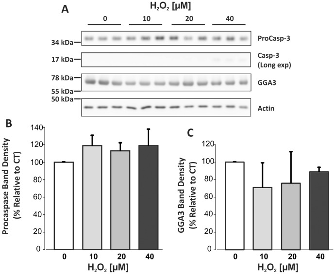
Figure 3. Immunoblot of caspase-3 and GGA3 in lysates of primary cortical cultures treated with H2O2.
20 µg of lysate was loaded per lane. A. Representative blots of caspase-3 and GGA3 from three experiments. Densitometry analyses of full-length proCaspase-3 35 kDa signal. C. Densitometry analysis of 75 kDa GGA3 signal. For both caspase-3 and GGA3, data were normalised to actin. One-way ANOVA indicated no significant change in levels of either protein in response to H2O2 treatments.

