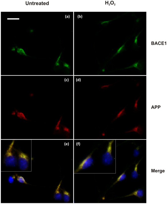
Figure 6. Immunofluorescence analysis of BACE1 and APP colocalisation. in mouse primary cortical neurons treated or untreated with H2O2.
Immunolabelling of untreated cells (a,c,e) and H2O2-treated cells (B). BACE1 was labelled with mouse antibody 61-3E7 (a,b) and APP was revealed with rabbit antibody 369 (c,d). Since Ab 369 targets the cytosolic domain of APP, it can stain both APP full-length and C-terminal fragments. Colocalisation of BACE1 and APP was observed in untreated cells (e) and this colocalisation may be increased in the treated cells (f). Bar = 20 µm.

