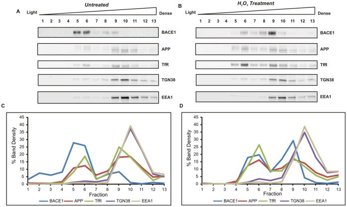
Figure 7. Subcellular fractionation of mouse primary cortical cells on a discontinuous sucrose density gradient.
Cells were incubated for 6 h in the presence or absence of 40 µM H2O2 and lysed under isotonic conditions. Post-nuclear supernatants were centrifuged at 100,000×g on a discontinuous sucrose gradient. Fractions were analysed by immunoblotting with antibodies for BACE1 (D10E5), APP (22C11) and for organelle markers (Transferrin receptor, TfR for plasma membrane and endocytic vesicles; TGN38, for trans-Golgi; EEA1 for early endosomes). Signal density was determined for all fractions, and specific protein levels in each fraction were expressed as a percentage of the total signals from all thirteen fractions. Representative blots from two experiments with untreated cells (A) and H2O2-treated cells (B). Graphs (C and D) represent signal intensity % of each protein plotted against fraction number. Noticeable changes in BACE1 distribution were observed after H2O2-treatment, with BACE1 protein redistributing from light to denser fractions, enriched in EEA1 and TGN38 markers.

