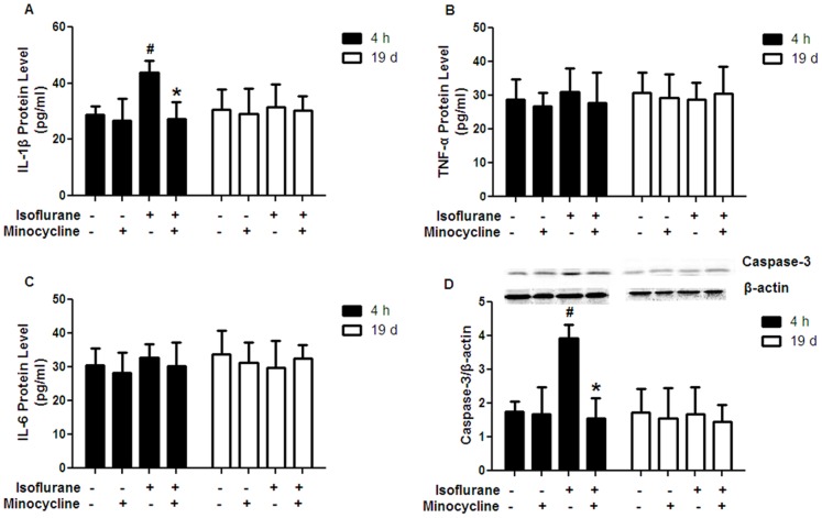
Figure 3. Effects of minocycline pretreatment on the levels of IL-1β, TNF-α, IL-6 and caspase-3 in the hippocampus.
(A) Minocycline application decreased the level of IL-1β at 4 h after isoflurane exposure, but not at 19 d after isoflurane exposure. (B) No differences were observed in TNF-α level among the four groups at 4 h or 19 d after isoflurane exposure. (C) No differences were observed in IL-6 level among the four groups at 4 h or 19 d after isoflurane exposure. (D) Minocycline application decreased the protein level of caspase-3 at 4 h after isoflurane exposure, but not at 19 d after isoflurane exposure. A representative Western blot is shown and the quantified caspase-3 bands were normalized to the loading control β-actin. 4 h = 4 h after isoflurane exposure; 19 d = 19 days after isoflurane exposure. Data represent mean ± S.E.M. n = 5 in each group. # P<0.05 isoflurane group compared with control group; *P<0.05 minocycline-isoflurane group compared with isoflurane group.

