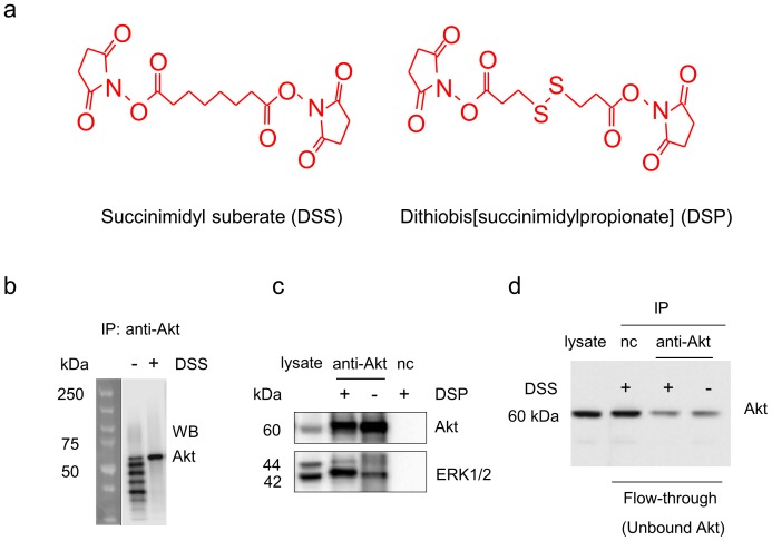
Figure 2. Western blot analysis of Akt and ERK1/2 monitored during co-IP procedures.
a. Chemical structures of the crosslinkers used in the study. b. Antibody contamination was eliminated by DSS-crosslinking of Akt antibody to protein A/G agarose beads. c. ERK1/2 co-immunoprecipitation with Akt was significantly improved by DSP-crosslinking. d. DSS-crosslinking did not alter antibody-antigen binding. nc, negative control using IgG instead of Akt antibody at a similar protein level.

