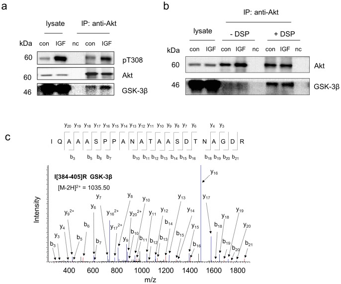
Figure 5. IGF-dependent interaction of Akt with GSK-3β revealed by the crosslinking/co-IP approach.
a, Western blot analysis indicating that active Akt is associated with GSK-3β more than inactive Akt. The active Akt status was indicated by phosphorylation of T308. b, Western blot analysis of the co-IP complex with or without DSP-crosslinking following the incubation of the cell lysate with Akt antibody immobilized on the beads by DSS. GSK-3β was detected in either control or IGF-stimulated samples only when DSP-crosslinking was used. c, Representative MS/MS spectrum of a peptide corresponding to GSK-3β identified only with DSP-crosslinking approach. The sequence of the peptide was assigned with a single letter abbreviation based on the fragment ions observed for the peptide segments. N-terminal b ions and C-terminal y ions resulting from the amide bond cleavage are labeled. Con, control sample without IGF stimulation. IGF, IGF-stimulated samples obtained by treating Neuro 2A cells with IGF at 10 ng/mL for 10 min. nc, negative control using IgG instead of Akt antibody at a similar protein level. * denotes the addition of 145.0198 Dalton due DSP-crosslinking.

