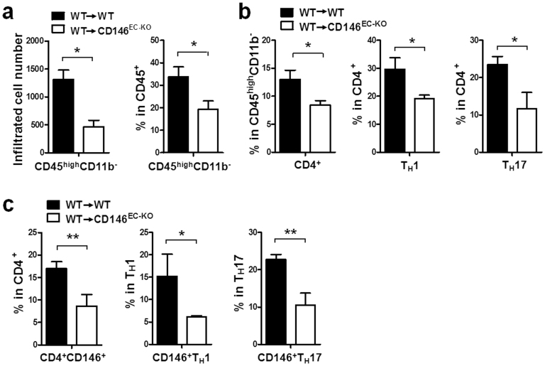
Figure 3. Fewer lymphocytes infiltrate the CNS in CD146EC-KO mice than in WT mice following the induction of passive EAE.
Flow cytometry analysis of CD45highCD11b− lymphocytes (a), CD4+ TH1 and TH17 lymphocytes (b) and CD146+ T cells (c) from the CNS of CD146EC-KO and WT mice with passive EAE. The data represent three independent experiments; 5 mice/group. *, p < 0.05, **, p < 0.01.

