Figure 1.
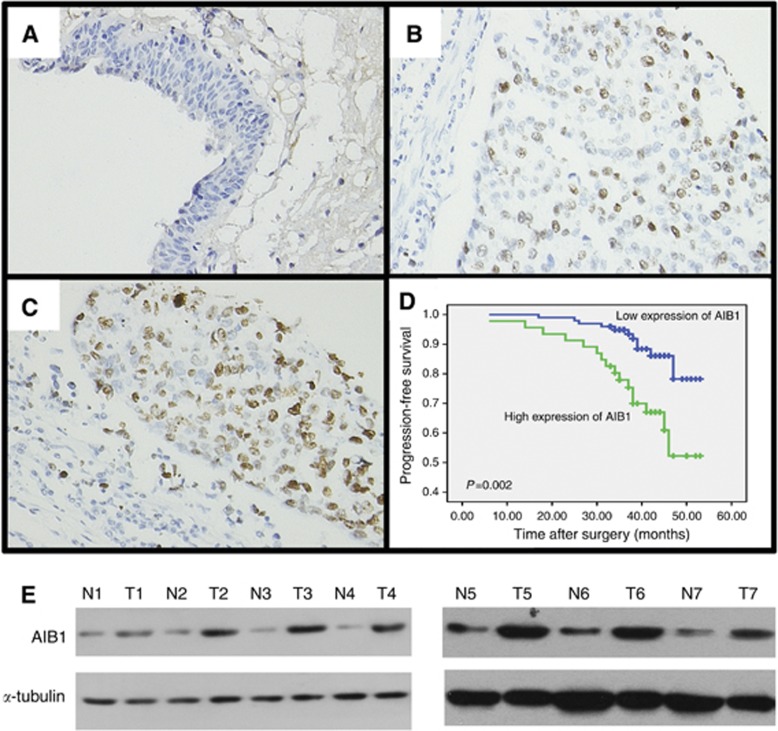
Expression and prognostic significance of amplified in breast cancer (AIB1) in BC. (A) Representative image of negative AIB1 IHC staining in normal bladder tissues. (B) Representative image of weak AIB1 IHC staining in BC tissues. (C) Representative image of positive AIB1 IHC staining in BC tissues. (D) Kaplan–Meier survival analysis according to AIB1 expression in 146 patients with non-muscle-invasive BC (log-rank test). (E) Western blotting analysis of AIB1 protein expression in pairs of matched BC (T) and adjacent normal tissues (N).

