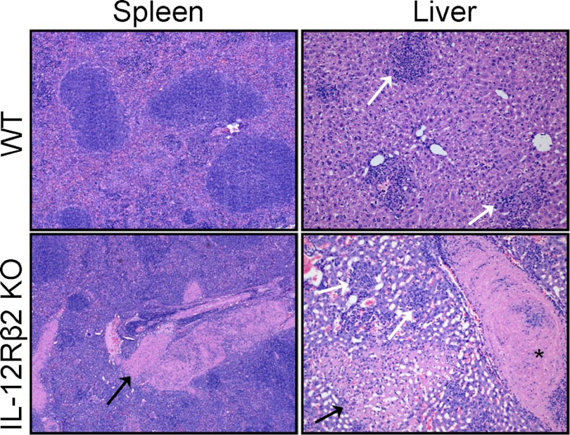
Figure 4. Representative organ sections from WT and IL-12Rβ2 KO mice.
WT and IL-12Rβ2 KO mice were infected with 105 LVS i.d. Spleens and livers were removed from two to three mice/group on Day 7 after infection, preserved in 10% formalin, embedded in paraffin, sectioned for slides, and stained with H&E. Black arrows indicate areas of necrosis; white arrows show granulomatous inflammation. *Large thrombus in hepatic central vein. These data are representative of two independent experiments of similar design.

