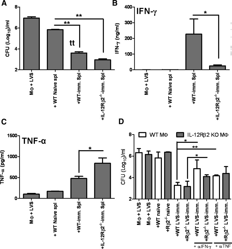
Figure 5. Control of F. tularensis LVS intracellular replication by LVS-immune lymphocytes.
BMDMs from WT mice were infected with F. tularensis LVS, and splenocytes from WT naïve mice or the indicated WT or IL-12Rβ2 KO LVS-immune (imm.) mice were added. Immune mice were primed with 103 LVS i.d. At 72 h after initiation of cocultures, (A) Macrophage lysates plated to enumerate bacteria, and supernatants were removed and assayed for levels of (B) IFN-γ and (C) TNF-α. Results shown are the mean CFU/ml ± sd of triplicate wells or mean ng/ml or pg/ml cytokine ± sd, and are representative of four independent experiments. (D) BMDMs derived from WT or IL-12Rβ2 KO mice were infected with F. tularensis LVS and cocultured with indicated splenocytes, and murine IFN-γ- or TNF-α-neutralizing antibodies (αIFN-γ and αTNF; 25 μg/ml) were added to the indicated wells. Bacterial growth was assessed 72 h after infection. Results shown are the mean CFU/ml ± sd of triplicate wells of one experiment and are representative of two independent experiments. *P ≤ 0.05; **P ≤ 0.01 by Student's t-test in a pair-wise comparison of samples from WT and KO mice.

