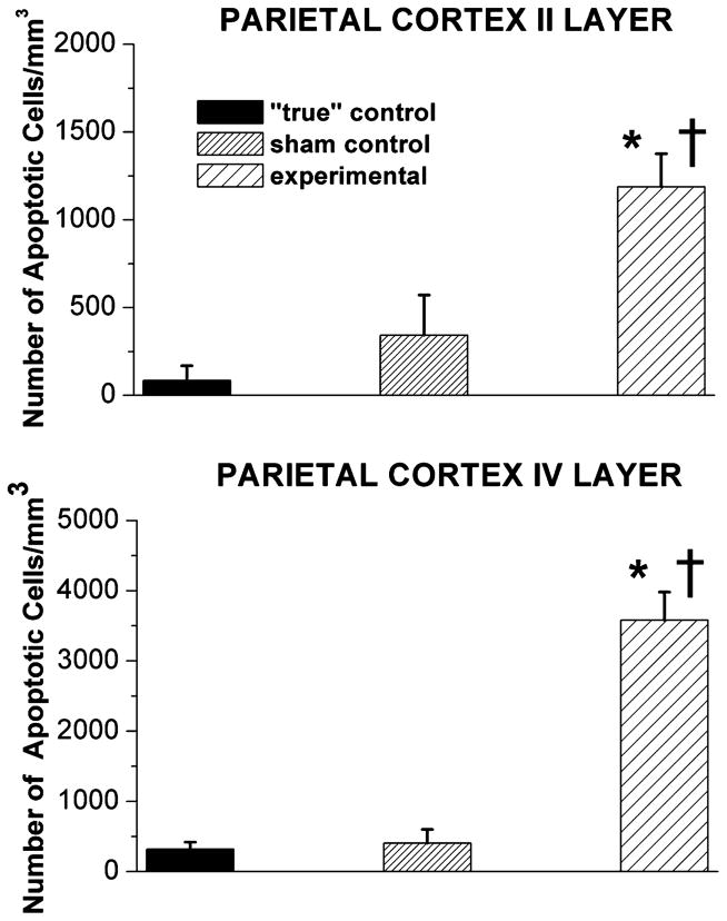
Figure 2.
Anesthesia induces significant caspase-3 activation in the developing parietal cortex of 5-day-old piglets. In piglet brains, the sensitivity of the parietal cortices to general anesthesia is quantified as the number of caspase-3-positive cells/mm3 in layers II and IV. The density of caspase-3-positive neurons in “true” control was minimal in parietal cortical regions. In comparison, although a somewhat higher degree of caspase-3 labeling occurred in sham control (exposed to a continuous fentanyl infusion, 15 μg/kg/h), the difference was not statistically significant. The combination of isoflurane (0.55-vol%), N2O (75 vol-%), and midazolam (1 mg/kg, i.m.) induced a significant increase in caspase-3 labeling as compared to labeling in both “true” and sham controls (*, P < 0.05, and †, P < 0.05, respectively in layer II; *, P < 0.001, and †, P < 0.001, respectively in layer IV) (n = 1 piglet per condition, n = 5 sections per brain region).

