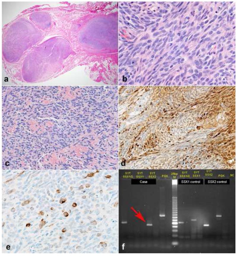
Figure 15. Intraneural synovial sarcoma mimicking malignant peripheral nerve sheath tumor (MPNST).
Synovial sarcoma represents the main entity in the differential diagnosis with MPNST, and similarly may infiltrate numerous nerve fascicles (a). Monotonous spindle cells in fascicles are characteristic (b) as well as variable intratumoral collagen bundles (c). Overt S100 immunoreactivity may be present in some synovial sarcomas (d). Cytokeratin typically labels isolated cells (e). Molecular confirmation may be required in some instances. In this example, RT-PCR reveals a SYT-SSX2 fusion transcript (arrow)(f), which is diagnostic of synovial sarcoma.

