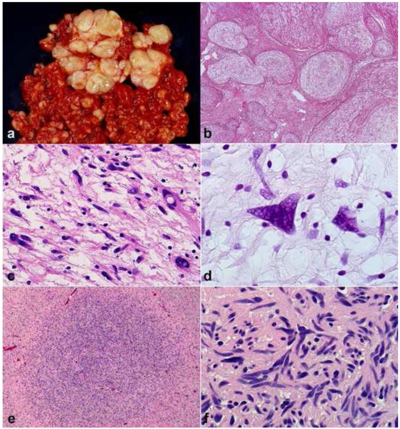
Figure 2. Diagnostically relevant neurofibroma variants.
Plexiform neurofibromas typically form large, multinodular masses (a). Involvement and expansion of multiple peripheral nerve fascicles is definitional (b). Degenerative atypia, in the absence of hypercellularity has been interpreted by some authors as “atypical neurofibroma”, has no prognostic significance, but may create diagnostic difficulties (c). The atypia in some neurofibromas is reflected in hyperchromatic nuclei with “smudgy” chromatin, which is probably degenerative (d). Foci of hypercellularity may be present in some neurofibromas (e). Areas of hypercellularity (“cellular” neurofibromas) in the absence of other atypical features is still compatible with a benign course (f).

