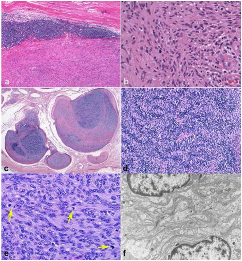
Figure 5. Important schwannoma variants.
Cellular schwannomas, despite their alarming cellularity usually have a well formed capsule, often with multiple subcapsular lymphoid aggregates (a). Verocay bodies are usually lacking (b). Plexiform schwannomas involving major peripheral nerves are rare, characteristically involving multiple fascicles (c). Plexiform schwannomas are composed primarily of Antoni A areas (d). Mitotic activity in plexiform/cellular schwannomas may be brisk (arrows), a finding still compatible with a benign diagnosis(e). Electron microscopy demonstrates extensive basal lamina in all schwannoma subtypes, a diagnostically useful finding (f).

