Figure 1.
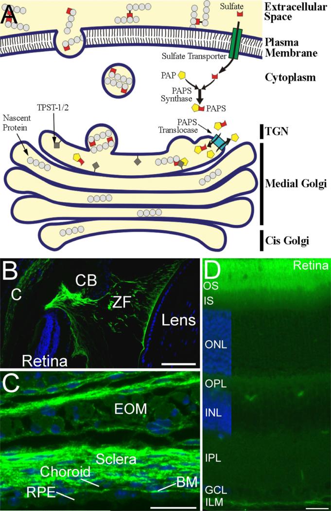
A. Figure 1. Synthetic pathway for generation of sulfotyrosine and localization of sulfotyrosine proteins in mouse eye. A. The enzymatic generation of sulfotyrosine. Sulfate donor 3'-phosphoadenosine 5'¬phosphosulfate (PAPS) is translocated into the trans-Golgi network (TGN) by the enzyme PAPS translocase from the cytoplasm where it is synthesized. Once inside the TGN, TPST-1/2 located in the TGN membrane incorporates the sulfate residue from PAPS into tyrosine residues of secreted and transmembrane proteins. The sulfotyrosine-containing proteins are then incorporated into vesicles to be transported to their final destination, which may either be the plasma membrane or the extracellular space. B-D. Localization of tyrosine sulfated proteins in ocular tissues. Sections were stained with anti-sulfotyrosine antibody (green) and the cell nuclei were counterstained with DAPI (blue). B. Tyrosine sulfated proteins are present in the cornea (C), and in the zonule fibers (ZF) and their attachments to the ciliary body (CB) and the lens. C. Tyrosine sulfated proteins are abundant in the sclera, choroid, Bruch's membrane (BM), and the connective tissue associated with the extraocular muscles (EOM). The retinal pigmented epithelium (RPE) and skeletal muscle cells of the EOM also show labeling for tyrosine sulfated proteins, but at much lower levels than the surrounding connective tissues. D. Tyrosine sulfated proteins are present in all layers of the retina, but are most abundant in the interphotoreceptor matrix surrounding the photoreceptor outer segments (OS) and at the inner limiting membrane (ILM). IS, inner segments; ONL, outer nuclear layer; OPL, outer plexiform layer; INL, inner nuclear layer; IPL, inner plexiform layer; GCL, ganglion cell layer. Scale bars = 100 μm for B; 20 μm for C and D.

